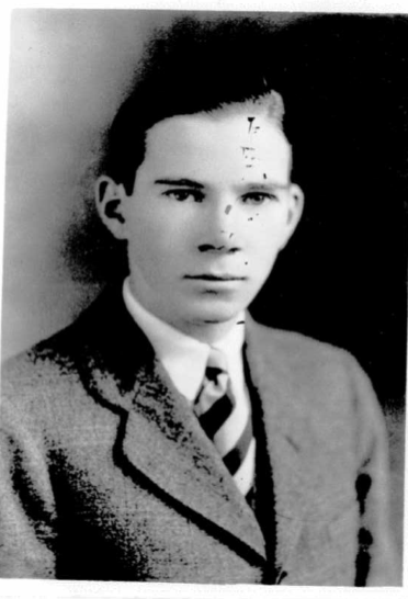
(11 July 1908 – 1987)
The University of North Carolina at Chapel Hill Herbarium (NCU) has cataloged vascular plant, bryological, and fungal specimens collected by Kenneth Bryan Raper, who usually signed his collections as “K.B. Raper.”
NCU’s fungal collection is cataloged at mycoportal.org , our moss, liverwort and hornwort collection is available via bryophyteportal.org , and our vascular plants are cataloged at sernecportal.org . Other herbaria curating collected by Kenneth B. Raper include the Farlow Herbarium of Harvard University (FH) and the US National Fungus Collection (BPI).
Kenneth Raper is best known for his work on Penicillium, the organism which produces the antibiotic Penicillin, as well as his work on slime molds. He discovered and described the slime mold Dictyostelium discoideum, which has become a model organism to study cell communication, differentiation, and programmed cell death. Kenneth Raper isolated D. discoideum “from decaying leaves collected in a hardwood forest of the North Carolina mountains in the summer of 1933. The dominant trees were beech [Fagus grandifolia], birch [Salix], oak [Quercus], and buckeye [Aesculus], and the sample consisted largely of the partially decomposed leaves of these trees…”8
—–
Kenneth Bryan Raper was born in Welcome, Davidson County, North Carolina on 11 July 1908 to Julia Salina (Crouse) Raper and William Franklin Raper, a farmer. He was one of eight children; three of his brothers also attended the University of North Carolina at Chapel Hill — Arthur F. (class of 1924, Ph.D. 1931), and Howard D. (class of 1927) and John R. (class of 1933).1,2
Kenneth Raper entered the University of North Carolina at Chapel Hill in 1925 and earned an A. B. in Botany in 1929. According to the “Dope Sheet” for the Class of 1929 in the 1979 Yackety-Yack, his nickname at Carolina was “Little Red” for the color of his hair, and to distinguish him from his brother, John “Red” Raper. The specimens in NCU’s collections were collected during his undergraduate years, and frequent collecting locations included Carrboro, Chapel Hill and his hometown of Welcome.
“I remember Chapel Hill as a small town occupied in large part by the University – the current Methodist, Baptist and Episcopal churches were relatively new and most inspiring to a country boy from up-state. South Building was being remodeled and the old coffin-shaped Memorial Hall was still standing. The oak trees in the front campus were majestic and those between South & the New Library were mere saplings. Davie Hall (Botany & Zoology) was the center of my activities and I remember well such famous professors as Wm. C. Coker, H.V. “Froggy” Wilson, Collier Cobb and Archibald Henderson (with whom I had one course and was scared to death.) Most vividly I remember Profs. John N. Couch and Frank P. Graham with whom I had courses that significantly influenced my future work and my appreciation of our American Heritage.” 2
Raper continued his education at George Washington University (M.A., 1931) and Harvard University (M.A., 1935 and Ph.D., 1936). He received an Honorary Doctor of Science from University of North Carolina at Chapel Hill in 1961.2
Kenneth Raper married Louise Montgomery Williams, a native of Charleston, West Virginia and graduate of Marshall College in 1936, and they adopted a son, Charles Albert Raper, born 18 August 1926. 1
Raper was employed as a Junior Mycologist at the United States Dept. of Agriculture in Washington, D.C. in 1929, then as a Principal Mycologist in the Northern Regional Research Laboratory of that agency in Peoria, Illinois in 1939, where he was “in charge of and largely responsible for the development and maintenance of the laboratory’s culture collection, built up through his efforts from a few hundred specimens to more than 5,000 industrially-important molds. The collection is internationally known for its great variety of micro-organisms that are available alike for research and industrial use.” 3
Raper worked in penicillin production during World War II in lieu of military service.
“We set out with three goals in mind. First, to find a better nutrient for the mold that would give higher yields. Second, to produce penicillin in submerged cultures so we could grow it in big tanks. Third, to find a mold that would produce more penicillin than the strain that came from Prof. Alexander Fleming’s lab.”4
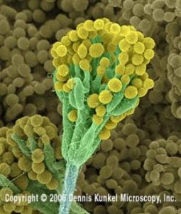
Mold conidiophores (fruiting structures/sporangia) and conidia (asexual spores) of
Penicillium chrysogenum
magnification 600x
Copyright Dennis Kunkel Microscopy, Inc.
www.denniskunkel.com
Image used with permission
In 1944 Raper and colleagues discovered Penicillium chrysogenum, which Raper called “the great grand-daddy” of all the cultures used to produce Penicillin, on a molded cantaloupe that a Peoria woman brought to the lab.5 While the original strain of Penicillium produced ca. 40 units/ml of penicillin, the Peoria cantaloupe strain produced 70-80 units/ml, and a mutant isolated from that strain yielded 250 units/ml. Industrial strains of P. chyrsogenum now produce 50,000 units/ml.6
In 1953 Raper joined the University of Wisconsin in Madison and by the time he retired from teaching in 1979, he was the William Trelease Professor of Bacteriology & Botany at that institution. While at Madison, Raper taught “Molds, Yeasts & Actinomyetes,” a course that detailed the natural history and commercial uses of mold from grains, fruits and soils.5 During the winter of 1974 Raper returned to Chapel Hill to study slime molds, a group that he’d studied as a graduate student. “I’m a dual personality in research. One part relates to mold which can be very, very practical, and the other to these slime molds, where, at the outset, I don’t think anyone saw any reason for studying them – except for the joy of finding out what was going on.”4
Raper was elected to Phi Beta Kappa at the University of North Carolina at Chapel Hill in 1929 and to the National Academy of Sciences in 1949. He was given the Charles Thow Award by the Society for Industrial Microbiology in 1966.
Raper died at age 79 in 1987, and his papers are curated at the LuEsther T. Mertz Library at the New York Botanical Garden.7
PUBLICATIONS (incomplete list)
1.
Title: Notice of Foray
Author(s): Kenneth B. Raper
Source: Mycologia, Vol. 42, No. 3 (May – Jun., 1950), p. 449
Publisher(s): Mycological Society of America
Stable URL: http://www.jstor.org/stable/3755799
2.
Title: Culture Collections of Microorganisms
Author(s): Kenneth B. Raper; R. E. Buchanan; P. R. Burkholder; R. E. Cleland; R. D. Coghill; John F. Enders; Carl Lamanna; E. H. Lennette; H. H. McKinney; Wm. J. Robbins; Joel Warren
Source: Science, New Series, Vol. 116, No. 3007 (Aug. 15, 1952), pp. 179-180
Publisher(s): American Association for the Advancement of Science
Stable URL: http://www.jstor.org/stable/1680095
3.
Title: Charles Thom: 1872-1956
Author(s): Kenneth B. Raper
Source: Mycologia, Vol. 49, No. 1 (Jan. – Feb., 1957), pp. 134-150
Publisher(s): Mycological Society of America
Stable URL: http://www.jstor.org/stable/3755740
4.
Title: The Yellow-Pigmented Dictyostelia
Author(s): James C. Cavender; Ann C. Worley; Kenneth B. Raper
Source: American Journal of Botany, Vol. 68, No. 3 (Mar., 1981), pp. 373-382
Publisher(s): Botanical Society of America
Stable URL: http://www.jstor.org/stable/2442773
Abstract: The cellular slime molds described herein are of a clear golden yellow color and belong to two naturally occurring groups. One we call Dictyostelium mexicanum sp. n. because it is found in soils from various parts of tropical and subtropical Mexico. The well-tapered stalk and disklike base are somewhat reminiscent of D. discoideum. The other is regarded as D. aureum E. W. Olive (1901) with which we feel it is closely identified if not identical. In the latter group the form of both the aggregation and sorocarp is suggestive of D. mucoroides. Some less pigmented isolates are separated as D. aureum var. luteolum var. n.
5.
Title: The Production and Characterization of Ultraviolet-Induced Mutations in Aspergillus terreus. III. Biochemical Characteristics of the Mutations
Author(s): Lewis B. Lockwood; Kenneth B. Raper; Andrew J. Moyer; Robert D. Coghill
Source: American Journal of Botany, Vol. 32, No. 4 (Apr., 1945), pp. 214-217
Publisher(s): Botanical Society of America
Stable URL: http://www.jstor.org/stable/2437512
Abstract: Nine different types of biochemical and cultural response have been observed from 217 strains of Aspergillus terreus derived from irradiated conidia. Among the 76 strains which were morphologically unchanged were 59 that appeared to be unaltered biochemically, 13 that produced more itaconic acid than the parent strain, and 4 that produced no itaconic acid. Among the 141 strains which were obviously altered morphologically were 42 strains not apparently altered biochemically, 88 which produced little acid, and 11 which did not grow on the test medium. None of these 141 strains produced more itaconic acid than did the parent strain. Fifteen strains produced considerable nonacidic unsaturated material. Seventeen strains appeared to produce no acid other than itaconic. The distribution of strains, plotted in terms of total acid produced, itaconic acid produced, efficiency of conversion of glucose to itaconic acid, mycelial weights, and neutral nonreducing materials produced, is presented.
6.
Title: The Production and Characterization of Ultraviolet-Induced Mutations in Aspergillus terreus. II. Cultural and Morphological Characteristics of the Mutations
Author(s): Kenneth B. Raper; Robert D. Coghill; Alexander Hollaender
Source: American Journal of Botany, Vol. 32, No. 3 (Mar., 1945), pp. 165-176
Publisher(s): Botanical Society of America
Stable URL: http://www.jstor.org/stable/2437538
Abstract: Conidia of a selected strain of Aspergillus terreus were exposed to ultraviolet radiation, and random isolations were made from colonies resulting from irradiated cells. Approximately two hundred of these were cultivated upon a variety of agar media to determine the effect of the substratum upon growth and attendant cultural characteristics. The majority of isolates from irradiated spores, normal appearing and cultural mutants alike, remained stable when recultivated through ten successive culture generations upon Czapek’s solution agar. Eleven types of mutations were recognized. One of these resulted from an inability of the mutant to produce thiamin. Another was unable to utilize nitrate nitrogen but developed normally if supplied with ammonia or amino nitrogen. In other cases mutations were more strictly morphological and consisted of a total or partial loss of color from the conidial heads without apparent alteration in physiology. In still other and more numerous cases, both morphological and physiological changes were observed. Cultural and morphological differences commonly reflected basic physiological or biochemical changes. Of more than 200 isolates resulting from irradiated spores tested for the production of itaconic acid, only 14 produced yields greater than the parent strain. Among this number only two appeared as cultural mutants, and only one of these was markedly different from the parent strain. The production of ultraviolet-induced mutations appears to offer definite, if somewhat limited, possibilities of increasing yields of itaconic acid from Aspergillus terreus.
7.
Title: The Production and Characterization of Ultraviolet-Induced Mutations in Aspergillus terreus. I. Production of the Mutations
Author(s): Alexander Hollaender; Kenneth B. Raper; Robert D. Coghill
Source: American Journal of Botany, Vol. 32, No. 3 (Mar., 1945), pp. 160-165
Publisher(s): Botanical Society of America
Stable URL: http://www.jstor.org/stable/2437537
Abstract: The method for the production of mutations in Aspergillus terreus by ultraviolet radiation is described. The probability of producing “deficient” mutants through chemical changes and the occasional appearance of mutants having increased ability to produce certain compounds is pointed out. Results are given on the per cent of mutation and survival after exposure to λ 2280, 2650, and 2967 Å. The effect of secondary treatment after irradiation and the characteristic appearance of mutations after a relatively small number of spores are killed are reported.
8.
Title: Sexuality in the Cellular Slime Mold Dictyostelium giganteum
Author(s): Gregory W. Erdos; Kenneth B. Raper; Linda K. Vogen
Source: Proceedings of the National Academy of Sciences of the United States of America, Vol. 72, No. 3 (Mar., 1975), pp. 970-973
Publisher(s): National Academy of Sciences
Stable URL: http://www.jstor.org/stable/64398
Abstract: By pairing of strains of Dictyostelium giganteum in various combinations this species was shown to be heterothallic. Four mating types were identified. Some strains could not be assigned a mating type and others showed no mating reaction. No self-compatible strains were found. Mutations were introduced in several strains and genetic crosses were performed. The results of these crosses show that mating and macrocyst formation are controlled by a single locus-multiple allele incompatibility system. The results also support the view that the myxamoebae that emerge upon germination of the macrocysts are the products of meiosis.
9.
Title: Mating Types and Macrocyst Formation in Dictyostelium discoideum
Author(s): Gregory W. Erdos; Kenneth B. Raper; Linda K. Vogen
Source: Proceedings of the National Academy of Sciences of the United States of America, Vol. 70, No. 6 (Jun., 1973), pp. 1828-1830
Publisher(s): National Academy of Sciences
Stable URL: http://www.jstor.org/stable/62229
Abstract: Strains of Dictyostelium discoideum were paired in various combinations. Certain pairings resulted in the production of macrocysts, which are thought to be the sexual stage in the cellular slime molds. One self-compatible strain was found. Other strains produced macrocysts only when paired with an alternate mating type. Two strains were found to form macrocysts when paired with either mating type but not alone or with each other. Several strains did not form macrocysts under any circumstances. Strains were graded according to their mating competency based on the number of macrocysts produced relative to the aggregated myxamoebae observed in the pairings.
10.
Title: Dictyostelium deminutivum, a New Cellular Slime Mold
Author(s): Johanna S. Anderson; Dorothy I. Fennell; Kenneth B. Raper
Source: Mycologia, Vol. 60, No. 1 (Jan. – Feb., 1968), pp. 49-64
Publisher(s): Mycological Society of America
Stable URL: http://www.jstor.org/stable/3757313
Abstract: A new species of Dictyostelium, isolated from leaf mold collected in Poza Rica, Mexico, is described. The slime mold is characterized by the relatively small dimensions of its spores and myxamoebae, and more especially by its diminutive sorocarps-hence the name D. deminutivum. The slime mold may be cultivated satisfactorily in association with either Escherichia coli or Pseudomonas fluorescens upon agar media of very low nutrient content. Optimal growth and development occur in association with the latter bacterium upon 0.025% glucose-0.025% peptone agar when glass Petri plate lids are replaced with porous clay covers at the time cell aggregation begins. Aggregations that subsequently produce normal sorocarps typically appear mound-like and arise by the concerted influx of unaligned myxamoebae, either singly or in small groups, rather than by the formation and convergence of definite streams. Depending upon their size, such pseudoplasmodia may occasionally yield single fructifications, but as a rule they produce several sorocarps which arise near the periphery of the cell mass. In the completed sorocarp, the sorophore tapers gradually from base to apex and consists uniformly of a single tier of cells, the outlines of which may be difficult to discern, particularly in the terminal area.
11.
Title: Dictyostelium aureo-stipes and Dictyostelium tenue: New Species of the Dictyosteliaceae
Author(s): James C. Cavender; Kenneth B. Raper; Ann M. Norberg
Source: American Journal of Botany, Vol. 66, No. 2 (Feb., 1979), pp. 207-217
Publisher(s): Botanical Society of America
Stable URL: http://www.jstor.org/stable/2442526
Abstract: Three new cellular slime molds, Dictyostelium aureo-stipes sp. n., D. aureo-stipes var. helvetium var. n. and D. tenue sp. n., are described which possess characteristics heretofore unrecorded in the Dictyosteliaceae. The two species are unlike in dimensions and complexity of form, yet show a number of features in common, and may in fact be closely related D. aureo-stipes var. helvetium is relatively large and robust, forming multiple-branched fruiting bodies without the regularity of form found in Polysphondylium, yet tending toward symmetry when well-developed. The golden-yellow stipe is a distinguishing feature of D. aureo-stipes and is even more pronounced in var. helvetium. D. tenue is smaller and simpler in form The degree of branching is much reduced, and oftentimes a solitary sorus terminates a delicate stipe composed of a single tier of cells. Both species are quite sensitive to environmental conditions, particularly temperature, for optimum development occurs within relatively narrow ranges.
12.
Title: Copromyxella: A New Genus of Acrasidae
Author(s): Kenneth B. Raper; Ann C. Worley; Terrence A. Kurzynski
Source: American Journal of Botany, Vol. 65, No. 9 (Oct., 1978), pp. 1011-1026
Publisher(s): Botanical Society of America
Stable URL: http://www.jstor.org/stable/2442688
Abstract: A new genus of cellular slime molds, Copromyxella, is described together with four species that comprise it Since it resembles Zopf’s Copromyxa more than any genus presently recognized, but differs from it in important characteristics such as the smaller dimensions of its cellular elements and the delicacy of its fructifications, the name Copromyxella is chosen for the new taxon Four species are included: C. silvatica, C. filamentosa, C. spicata, and C. coralloides. All are characterized by small myxamoebae with lobose and potentially “explosive” pseudopodia and may assume limax forms in liquid media; all possess nuclei with centrally positioned nucleoli; all lack true contractile vacuoles (with a single possible exception); all aggregate without stream formation; and all form fructifications with no demarcation into stalks and sori. Taxonomically the genus belongs in the Acrasidae with Copromyxa, Guttulinopsis and Acrasis rosea rather than in the better known Dictyostelidae that includes Dictyostelium and Polysphondylium.
13.
Title: Ultrastructural Aspects of Two Species of Guttulinopsis
Author(s): Gregory W. Erdos; Kenneth B. Raper
Source: American Journal of Botany, Vol. 65, No. 5 (May – Jun., 1978), pp. 552-561
Publisher(s): Botanical Society of America
Stable URL: http://www.jstor.org/stable/2442589
Abstract: Two acrasid cellular slime molds, Guttulinopsis vulgaris and G. nivea, are compared at the ultrastructural level. The amoebae of the two species are indistinguishable except for the presence of intranuclear fibers in G. vulgaris. Both species share some unusual features, including: plate-like cristae in the mitochondria, production of microbody-like organelles in the perinuclear space, spores with thin bilaminar walls, and stalks containing microfilaments bound in striated bundles These and other observations are discussed with regard to the development of the sorocarps and the relationship of the genus to other members of the Acrasida.
14.
Title: Two Noteworthy Fungi from Liberian Soil
Author(s): Kenneth B. Raper; Dorothy I. Fennell
Source: American Journal of Botany, Vol. 39, No. 1 (Jan., 1952), pp. 79-86
Publisher(s): Botanical Society of America
Stable URL: http://www.jstor.org/stable/2438097
Abstract: Two striking fungi have been isolated from Liberian forest soil. One of these is Heterocephalum aurantiacum Thaxter; the other represents a new genus and species of the Kickxellaceae, for which the name Linderina pennispora is proposed. Cultural and morphological characteristics of each fungus are presented, together with some discussion regarding their probable relationships.
15.
Title: Two New Aspergillus nidulans Group Members from Wyoming Soils
Author(s): Martha Christensen; Kenneth B. Raper; Jack S. States
Source: Mycologia, Vol. 70, No. 2 (Mar. – Apr., 1978), pp. 332-342
Publisher(s): Mycological Society of America
Stable URL: http://www.jstor.org/stable/3759031
Abstract: Two new species in the Aspergillus nidulans group are described and compared with known related taxa. Aspergillus spectabilis, with tall conidiophores, broad green conidial columns and early-ripening cleistothecia shallowly embedded in a vinaceous Hülle-cell crust, may be most closely related to A. multicolor, A. crustosus (both nonascosporic), A. striatus and Emericella desertorum. Aspergillus bicolor is distinctive among all other recognized species in the complex in its development of tall conidiophores and columnar green heads on dilute media and in its production on malt agar of a floccose yellow felt which forms vinaceous Hülle-cell-cleistothecium aggregations aerially after growth for 4-6 wk. Ascospores in the latter species are produced sparingly, mature at 2-3 mo, are orange red, smooth or delicately roughened and have very low equatorial crests.
16.
Title: The Occurrence and Distribution of Acrasieae in Forests of Subtropical and Tropical America
Author(s): James C. Cavender; Kenneth B. Raper
Source: American Journal of Botany, Vol. 55, No. 4 (Apr., 1968), pp. 504-513
Publisher(s): Botanical Society of America
Stable URL: http://www.jstor.org/stable/2440581
Abstract: Forests of the subtropical and tropical regions of North America harbor cellular slime molds not found in the soils of temperate deciduous forests investigated previously. However, most species found in the temperate forest are common in the tropics. Although the diversity of forms is greater in the soils of tropical forests the numbers of Acrasieae per unit of soil are comparable. Characteristic of tropical and subtropical forest soils are Acrasieae bearing crampon bases, of which four new species of Dictyostelium are presently known. Also present, but less frequently isolated, are two other new species of the genus Dictyostelium and two still undescribed species of the Guttulinaceae. Occasional isolates of D. purpureum and D. discoideum were found that produce macrocysts, which seem, also, to be confined to tropical and subtropical areas. Macrocysts were previously known only in D. mucoroides and D. minutum isolated from temperate forest soils. The occurrence and distribution of Acrasieae in warm temperate desert and mesquite-scrub, in subtropical hammock, and in tropical thorn, deciduous, seasonal evergreen, rain, and cloud forests were investigated. Acrasieae were well represented in all of these forests except desert. The number of species and the total populations were largest in seasonal evergreen forests. The composition of the cellular slime mold populations and the dominant species within these populations could be related to the soil environment as expressed by the dominant vegetation.
17.
Title: The Influence of Light on the Time of Cell Aggregation in the Dictyosteliaceae
Author(s): Theo M. Konijn; Kenneth B. Raper
Source: Biological Bulletin, Vol. 128, No. 3 (Jun., 1965), pp. 392-400
Publisher(s): Marine Biological Laboratory
Stable URL: http://www.jstor.org/stable/1539901
18.
Title: The Influence of Light on the Size of Aggregations in Dictyostelium discoideum
Author(s): Theo M. Konijn; Kenneth B. Raper
Source: Biological Bulletin, Vol. 131, No. 3 (Dec., 1966), pp. 446-456
Publisher(s): Marine Biological Laboratory
Stable URL: http://www.jstor.org/stable/1539984
19.
Title: The Crampon-Based Dictyostelia
Author(s): Kenneth B. Raper; Dorothy I. Fennell
Source: American Journal of Botany, Vol. 54, No. 5 (May – Jun., 1967), pp. 515-528
Publisher(s): Botanical Society of America
Stable URL: http://www.jstor.org/stable/2440655
Abstract: Four new species of Dictyostelium, all with sorophore bases of a unique crampon-like pattern, are recognized among 25 isolates obtained from samples of soil and leaf litter collected in forests of Panama, Mexico, Costa Rica, Florida, and Texas. The species described are as follows: D. rhizopodium, characterized by frequently clustered sorocarps with yellowish to grayish olive sori and yellow or brownish purple sorophores; D. coeruleo-stipes with white or cream-colored sori and purplish blue sorophores; D. lavandulum with grayish lavender to drab vinaceous sori and vinaceous purple sorophores; and D. vinaceo-fuscum with deep vinaceous gray to dark smoke-colored sori and lavender sorophores.
20.
Title: The Acrasieae in Nature. III. Occurrence and Distribution in Forests of Eastern North America
Author(s): James C. Cavender; Kenneth B. Raper
Source: American Journal of Botany, Vol. 52, No. 3 (Mar., 1965), pp. 302-308
Publisher(s): Botanical Society of America
Stable URL: http://www.jstor.org/stable/2439945
Abstract: The distribution of 9 members of the Acrasieae in the climax associations of the temperate deciduous forest of Eastern North America was determined. No undescribed species were isolated. The presence, density, relative density, and frequency of species were found to fluctuate with the environment as indicated by the dominant trees in the forest canopy. The acrasian population of the lowland hardwood forest (silver maple-American elm) differs markedly in relative density and frequency of its constituent species from that of any upland forest. High frequencies of Polysphondylium pallidum and Dictyostelium polycephalum characterize this forest type. In the upland forest, D. minutum is the most characteristic species and maintains a high frequency and relative density over most of the forest area; however, it decreases in importance in southern regions. The relatively dry oak-hickory association was found most favorable for the development of the Acrasieae. Soils of the boreal forest contain only limited numbers of Acrasieae representing few species.
21.
Title: The Acrasieae in Nature. II. Forest Soil as a Primary Habitat
Author(s): James C. Cavender; Kenneth B. Raper
Source: American Journal of Botany, Vol. 52, No. 3 (Mar., 1965), pp. 297-302
Publisher(s): Botanical Society of America
Stable URL: http://www.jstor.org/stable/2439944
Abstract: Although the Acrasieae are widely distributed in nature, they are most numerous and of the greatest variety in forest soils. Those species isolated from deciduous forest soils of Eastern and Midwestern United States belong to 2 families, the Dictyosteliaceae and Acytosteliaceae. Preference of the Acrasieae for forest soils over those of grassland was demonstrated by sampling a vegetational succession from prairie to forest. Occurrence of these slime molds within the forest soil profile is largely confined to the surface of the humus layer and the fermenting leaf litter. The Acrasieae are more numerous in forest soils in the autumn and spring than at other seasons. The frequency of occurrence of species varies considerably along the forest continuum of southern Wisconsin, indicating a differential response to microenvironments as influenced by the higher vegetation.
22.
Title: The Acrasieae in Nature. I. Isolation
Author(s): James C. Cavender; Kenneth B. Raper
Source: American Journal of Botany, Vol. 52, No. 3 (Mar., 1965), pp. 294-296
Publisher(s): Botanical Society of America
Stable URL: http://www.jstor.org/stable/2439943
Abstract: The clonal isolation of Acrasieae from suspensions of soils or organic substrates may be accomplished by employing a dilute hay-infusion agar medium to the surface of which pregrown bacteria have been added as a source of nutrient for the myxamoebae. The method described permits quantitative sampling of cellular slime mold populations, and it has been used by the authors in a study of the occurrence and distribution of these organisms in North and Central American forests.
23.
Title: Stalk Formation in Dictyostelium
Author(s): Kenneth B. Raper; Dorothy I. Fennell
Source: Bulletin of the Torrey Botanical Club, Vol. 79, No. 1 (Jan. – Feb., 1952), pp. 25-51
Publisher(s): Torrey Botanical Society
Stable URL: http://www.jstor.org/stable/2482103
Abstract: 1. Sorocarps, or fruiting structures, in Dictyostelium are formed by the orderly differentiation of large numbers of separate but clearly coordinated amoeboid cells, or myxamoebae. 2. The sorophore, or stalk, consists of an outer sheath of cellulose surrounding a column of closely compacted, strongly vacuolated, pith-like cells. The sheath appears in advance of obvious cell differentiation and is formed by the extracellular deposition of cellulose in a critical circular zone, the dimensions of which are proportional to the mass of cooperating myxamoebae. 3. The principal force responsible for lifting the sorogen, or sporogenous mass, into the air results from the swelling of stalk cells entrapped in the elongating sorophore sheath; a second and progressively diminishing force results from the coordinated pseudopodial movements of the myxamoebae that comprise the sorogen. Tests which indicate that the sorophore sheath (of extracellular origin) is cellulosic in character include: staining reactions, solubilities, birefringence in polarized light, X-ray diffracton pattern, paper chromatography of hydrolysis products, and decomposition by cellulose- destroying bacteria.
24.
Title: Spore Germination in Dictyostelium discoideum
Author(s): David A. Cotter; Kenneth B. Raper
Source: Proceedings of the National Academy of Sciences of the United States of America, Vol. 56, No. 3 (Sep. 15, 1966), pp. 880-887
Publisher(s): National Academy of Sciences
Stable URL: http://www.jstor.org/stable/57669
25.
Title: Observations on Guttulinopsis vulgaris and Guttulinopsis nivea
Author(s): Kenneth B. Raper; Ann C. Worley; Dietrich Kessler
Source: Mycologia, Vol. 69, No. 5 (Sep. – Oct., 1977), pp. 1016-1030
Publisher(s): Mycological Society of America
Stable URL: http://www.jstor.org/stable/3758784
Abstract: The genus Guttulinopsis of E. W. Olive is briefly reviewed and the common coprophilous species G. vulgaris is considered with regard to its distinguishing characteristics, the development of its fructifications (sorocarps) and its cultivation in the laboratory upon sterile dung partially embedded in low-nutrient agar. More detailed attention is given to the new species G. nivea that differs from the older species in important particulars: it produces sorocarps with well defined cellular stalks (sorophores) of a characteristic tapered pattern; it bears globose to subglobose spore heads (sori) that are consistently snow white when young, becoming lightly pigmented with age; and it can be cultivated quite successfully in two-membered cultures with either Klebsiella pneumoniae or Escherichia coli upon relatively weak glucose-peptone agar buffered to slightly acid pH. The myxamoebae of G. nivea, like those of G. vulgaris have lobose pseudopodia, nuclei (usually single) with centrally positioned nucleoli, usually single contractile vacuoles, and spores that often show prominent crescent-shaped vacuolation situated just inside the spore wall. Cell aggregation in G. nivea leading to sorocarp formation, as in G. vulgaris, occurs without stream formation, the converging myxamoebae responding individually to some chemotactic attractant of unknown nature.
26.
Title: Non-Viability of Stalk Cells in Dictyostelium
Author(s): William F. Whittingham; Kenneth B. Raper
Source: Proceedings of the National Academy of Sciences of the United States of America, Vol. 46, No. 5 (May 15, 1960), pp. 642-649
Publisher(s): National Academy of Sciences
Stable URL: http://www.jstor.org/stable/70897
27.
Title: Macrocysts in the Life Cycle of the Dictyosteliaceae. II. Germination of the Macrocysts
Author(s): Ann Weinkauff Nickerson; Kenneth B. Raper
Source: American Journal of Botany, Vol. 60, No. 3 (Mar., 1973), pp. 247-254
Publisher(s): Botanical Society of America
Stable URL: http://www.jstor.org/stable/2441215
Abstract: Macrocyst germination was demonstrated in the five species of the Dictyosteliaceae known to produce these structures. The morphological changes that occurred during germination appeared to be identical in all of the strains examined, showing the following stages: (1) swelling of the dark, contracted content of the dormant cysts, (2) gradual loss of color and reappearance of cells within what previously appeared as a homogeneous protoplasmic mass, and (3) rupture of the heavy cellulosic cyst wall to liberate the myxamoebae. The age of the macrocyst appeared to be the most critical factor in determining whether or not germination would occur, since the cysts in many of the strains needed to age for several weeks or months before germination could be demonstrated. In Dictyostelium mucoroides strain DM-7, upon which the current study was centered, light was necessary to stimulate germination of young macrocysts-a requirement that gradually diminished as the cysts aged. The rate of germination and the temperature permitting germination were also age dependent: older macrocysts germinated more rapidly and at considerably higher temperatures than did young cysts. Although light was not essential for germination in every strain, the results obtained with strain DM-7 seem to be generally applicable to the germination process.
28.
Title: Macrocysts in the Life Cycle of the Dictyosteliaceae. I. Formation of the Macrocysts
Author(s): Ann Weinkauff Nickerson; Kenneth B. Raper
Source: American Journal of Botany, Vol. 60, No. 2 (Feb., 1973), pp. 190-197
Publisher(s): Botanical Society of America
Stable URL: http://www.jstor.org/stable/2441106
Abstract: Macrocysts, a morphogenetic phase that is alternative to sorocarp formation in the life cycle of some cellular slime molds, are known for two genera and five species of the Dictyosteliaceae. In all of these macrocyst formation was found to be strongly affected by four factors: light, temperature, moisture, and the composition of the medium. In general, macrocyst formation was suppressed and sorocarp formation was enhanced by exposure to light, by incubation at temperatures lower than 20 C, by buffering nutrient media with phosphates, and by reducing atmospheric moisture through the use of clay covers on Petri dishes. The extent to which these environmental factors, singly or in combination, inhibited the production of macrocysts varied among the different strains and species.
29.
Title: Inhibition of Normal Pigment Synthesis in Spores of Dictyostelium purpureum
Author(s): William F. Whittingham; Kenneth B. Raper
Source: American Journal of Botany, Vol. 43, No. 9 (Nov., 1956), pp. 703-708
Publisher(s): Botanical Society of America
Stable URL: http://www.jstor.org/stable/2438836
Abstract: Phenol concentrations of 10-2 M to 5 x 10⁻¹ M induced the formation of white sori in cultures of Dictyostelium purpureum. This change in sorus color could not be attributed to a pH phenomenon, but rather, it is believed to result from an inhibition of the enzymatic sequence in pigment synthesis. Three additional compounds, m-cresol, p-cresol, and resorcinol, were found to induce effects similar to those induced by phenol. The minimum concentrations required to duplicate these effects were the same for all four compounds, suggesting that their modes of action were similar, if not identical. The relationship of this phenomenon to speciation in the Acrasieae is also discussed.
30.
Title: Further Investigations on the Preservation of Mold Cultures
Author(s): Dorothy I. Fennell; Kenneth B. Raper; May H. Flickinger
Source: Mycologia, Vol. 42, No. 1 (Jan. – Feb., 1950), pp. 135-147
Publisher(s): Mycological Society of America
Stable URL: http://www.jstor.org/stable/3755246
31.
Title: Fonticula alba: A New Cellular Slime Mold (Acrasiomycetes)
Author(s): Ann C. Worley; Kenneth B. Raper; Marianne Hohl
Source: Mycologia, Vol. 71, No. 4 (Jul. – Aug., 1979), pp. 746-760
Publisher(s): Mycological Society of America
Stable URL: http://www.jstor.org/stable/3759186
Abstract: Fonticula alba fits no presently recognized genus of the Acrasiomycetes. The slime mold grows and fruits well at 25-28 C with Klebsiella pneumoniae upon weakly buffered nutrient agar. The myxamoebae are small, variable in form and dimensions, uninucleate or less commonly binucleate and with nucleoli indistinct. Myxamoebae form no streams but aggregate along definite paths to form low rounded mounds. From these mounds narrow tapering columns of cells develop vertically, up to 1.0 mm, each bounded by a thin, tube-like covering that is closed above and extends downward over a broader basal reservoir of cells. Later the covering ruptures apically and the cells within ascend, as if projected under pressure, and collect into a spherical droplet to differentiate as spores. When mature, the naked sorus thus formed may rest upon the emptied hyaline tube for several da before the structure collapses. In no other cellular slime mold are sorocarps known to be formed in a similar manner. Myxamoebae that fail to aggregate differentiate individually as microcysts.
32.
Title: Environmental Factors Influencing the Growth and Fructification of Dictyostelium polycephalum
Author(s): William F. Whittingham; Kenneth B. Raper
Source: American Journal of Botany, Vol. 44, No. 7 (Jul., 1957), pp. 619-627
Publisher(s): Botanical Society of America
Stable URL: http://www.jstor.org/stable/2438936
Abstract: The relative humidity within the culture vessel, or the gradient between the atmosphere of the vessel and the external atmosphere, was of primary importance in obtaining optimum fruiting of Dictyostelium polycephalum. Proper control of this environmental factor permitted the slime mold to complete its life cycle with optimum formation of sorocarps in the absence of a third cultural associate, Dematium nigrum, which had previously been required. Apparently, the relative humidity required to induce the formation of sorocarps is slightly lower for D. polycephalum than for other members of the genus. In addition, proper manipulation of the culture atmosphere reduced the time required for maximum sorocarp production from 10-21 days to 5-6 days. Optimum fruiting was also dependent upon the pH of the substrate, the minimum being 5.6, the maximum 8.7, and the optimum 6.5. Therefore, the range within which sorocarps will differentiate appears to be shifted toward the basic side when compared to the ranges reported for most of the other Acrasieae. The optimum temperature for sorocarp formation and growth was found to be 30⚬C. while the minimum temperature for fruiting, but not growth, was 10-15⚬C. The nutrition of the bacterial associate was also instrumental in affecting the fruiting process of the slime mold; e.g., it was shown that the acidic and basic end-products in the metabolism of Escherichia coli interfered with sorocarp formation. This inhibition was not caused by the products per se, but rather resulted from the adverse pH which they induced in the substrate. If the bacterial growth was restricted, the pH of the substrate remained within a favorable range, and near the optimum, for fruiting. These results indicate that the primary beneficial function of Dematium in the culture of D. polycephalum, as previously reported by Raper, was the reduction of the humidity within the culture vessel which stimulated the differentiation of pseudoplasmodia into mature sorocarps. A second function of the Dematium could have been the removal, or neutralization, of acidic or basic end-products resulting from the metabolism of the bacterial associate.
33.
Title: Encystment Stages of Dictyostelium
Author(s): Joan Cormier Blaskovics; Kenneth B. Raper
Source: Biological Bulletin, Vol. 113, No. 1 (Aug., 1957), pp. 58-88
Publisher(s): Marine Biological Laboratory
Stable URL: http://www.jstor.org/stable/1538802
34.
Title: A Theory of the Control of Differentiation in the Cellular Slime Molds
Author(s): John Tyler Bonner; Kenneth B. Raper
Source: The Quarterly Review of Biology, Vol. 51, 50th Anniversary Special Issue, 1926-1976 (1976), pp. 296-312
Publisher(s): The University of Chicago Press
Stable URL: http://www.jstor.org/stable/2823089
35.
Title: Ashbya gossypii: Its Significance in Nature and in the Laboratory
Author(s): Thomas G. Pridham; Kenneth B. Raper
Source: Mycologia, Vol. 42, No. 5 (Sep. – Oct., 1950), pp. 603-623
Publisher(s): Mycological Society of America
Stable URL: http://www.jstor.org/stable/3755395
36.
Title: The Golden Anniversary of the Centraal Bureau Voor Schimmelcultures: 1907-1957
Author(s): Kenneth B. Raper
Source: Mycologia, Vol. 49, No. 6 (Nov. – Dec., 1957), pp. 884-892
Publisher(s): Mycological Society of America
Stable URL: http://www.jstor.org/stable/3755811
37.
Title: The Communal Nature of the Fruiting Process in the Acrasieae
Author(s): Kenneth B. Raper
Source: American Journal of Botany, Vol. 27, No. 6 (Jun., 1940), pp. 436-448
Publisher(s): Botanical Society of America
Stable URL: http://www.jstor.org/stable/2436460
38.
Title: The Aspergillus nidulans Group
Author(s): Charles Thom; Kenneth B. Raper
Source: Mycologia, Vol. 31, No. 6 (Nov. – Dec., 1939), pp. 653-669
Publisher(s): Mycological Society of America
Stable URL: http://www.jstor.org/stable/3754337
39.
Title: The Ascosporic Stage of Aspergillus citrisporus and Related Forms
Author(s): Kenneth B. Raper; Dorothy I. Fennell; Homer D. Tresner
Source: Mycologia, Vol. 45, No. 5 (Sep. – Oct., 1953), pp. 671-692
Publisher(s): Mycological Society of America
Stable URL: http://www.jstor.org/stable/4547748
40.
Title: The Arsenic Fungi of Gosio
Author(s): Charles Thom; Kenneth B. Raper
Source: Science, New Series, Vol. 76, No. 1980 (Dec. 9, 1932), pp. 548-550
Publisher(s): American Association for the Advancement of Science
Stable URL: http://www.jstor.org/stable/1656842
41.
Title: Studies on Variation and Mutation in Ashbya gossypii
Author(s): Thomas G. Pridham; Kenneth B. Raper
Source: Mycologia, Vol. 44, No. 4 (Jul. – Aug., 1952), pp. 452-469
Publisher(s): Mycological Society of America
Stable URL: http://www.jstor.org/stable/4547619
42.
Title: Sexuality and Cultural Characteristics of Aspergillus heterothallicus
Author(s): Kyung-Joo Kwon; Kenneth B. Raper
Source: American Journal of Botany, Vol. 54, No. 1 (Jan., 1967), pp. 36-48
Publisher(s): Botanical Society of America
Stable URL: http://www.jstor.org/stable/2440885
Abstract: Aspergillus heterothallicus K., F. and R., isolated from Costa Rican soils, represents the first species in this genus that is truly heterothallic. Each of the eleven strains investigated is functionally hermaphroditic but self-sterile and falls into one of two cross-mating classes, A or a. The mating-type factors A and a appear to be an allelic pair segregating independently of the locus for pigmentation of mycelium which varies from yellow to pinkish orange in different isolates. The striking variation in the development of hulle cell masses that occurred among the progeny from the cross WB 5096(A) x WB 5097(a) was found to be genetically controlled. The gene responsible for a delay in the formation and maturation of cleistothecia appeared to be loosely linked to the a mating-type locus and could be recombined into the A mating type. The mechanism of fertilization has not been completely elucidated. Coiled ascogonia were found within young hulle cell masses developed in cultures where two isolates of opposite mating types were crossed; such coils have not been observed, thus far, within hulle cell masses in unmated cultures. Although no recognizable male structure has been found, the fertilizing element appears to be mycelial in form rather than conidial. Interspecific mating did not occur when strains of Aspergillus heterothallicus were paired with other members of the A. nidulans group.
43.
Title: Preservation of Molds by the Lyophil Process
Author(s): Kenneth B. Raper; Dorothy F. Alexander
Source: Mycologia, Vol. 37, No. 4 (Jul. – Aug., 1945), pp. 499-525
Publisher(s): Mycological Society of America
Stable URL: http://www.jstor.org/stable/3754636
44.
Title: Nomenclature in Aspergillus and Penicillium
Author(s): Kenneth B. Raper
Source: Mycologia, Vol. 49, No. 5 (Sep. – Oct., 1957), pp. 644-662
Publisher(s): Mycological Society of America
Stable URL: http://www.jstor.org/stable/3755984
45.
Title: New Species of Penicillium
Author(s): Kenneth B. Raper; Dorothy I. Fennell
Source: Mycologia, Vol. 40, No. 5 (Sep. – Oct., 1948), pp. 507-546
Publisher(s): Mycological Society of America
Stable URL: http://www.jstor.org/stable/3755256
46.
Title: New Species and Varieties of Aspergillus
Author(s): Dorothy I. Fennell; Kenneth B. Raper
Source: Mycologia, Vol. 47, No. 1 (Jan. – Feb., 1955), pp. 68-89
Publisher(s): Mycological Society of America
Stable URL: http://www.jstor.org/stable/3755757
47.
Title: New Aspergilli from Soil
Author(s): Kenneth B. Raper; Charles Thom
Source: Mycologia, Vol. 36, No. 6 (Nov. – Dec., 1944), pp. 555-575
Publisher(s): Mycological Society of America
Stable URL: http://www.jstor.org/stable/3754836
48.
Title: Microbes — Man’s Mighty Midgets
Author(s): Kenneth B. Raper
Source: American Journal of Botany, Vol. 44, No. 1 (Jan., 1957), pp. 56-65
Publisher(s): Botanical Society of America
Stable URL: http://www.jstor.org/stable/2438345
49.
Title: Levels of Cellular Interaction in Amoeboid Populations
Author(s): Kenneth B. Raper
Source: Proceedings of the American Philosophical Society, Vol. 104, No. 6 (Dec. 15, 1960), pp. 579-604
Publisher(s): American Philosophical Society
Stable URL: http://www.jstor.org/stable/985535
50.
Title: Isolation, Cultivation, and Conservation of Simple Slime Molds
Author(s): Kenneth B. Raper
Source: The Quarterly Review of Biology, Vol. 26, No. 2 (Jun., 1951), pp. 169-190
Publisher(s): The University of Chicago Press
Stable URL: http://www.jstor.org/stable/2809212
51.
Title: Interspecific Mixtures in the Dictyosteliaceae
Author(s): Kenneth B. Raper; Charles Thom
Source: American Journal of Botany, Vol. 28, No. 1 (Jan., 1941), pp. 69-78
Publisher(s): Botanical Society of America
Stable URL: http://www.jstor.org/stable/2437063
Abstract: The western North American grass which Hitchcock had merged under the Chilean Poa secunda Presl is restored as a valid species, P. Sandbergii Vasey, and for it is given a new detailed description drawn from the type and from specimens from the vicinity of the type locality.
52.
Title: Homothallism vs. Heterothallism in the Penicillium luteum Series
Author(s): Kenneth B. Raper; Dorothy I. Fennell
Source: Mycologia, Vol. 44, No. 1 (Jan. – Feb., 1952), pp. 101-111
Publisher(s): Mycological Society of America
Stable URL: http://www.jstor.org/stable/4547570
53.
Title: Heterkaryon Formation and Genetic Analyses of Color Mutants in Aspergillus heterothallicus
Author(s): Kyung-Joo Kown; Kenneth B. Raper
Source: American Journal of Botany, Vol. 54, No. 1 (Jan., 1967), pp. 49-60
Publisher(s): Botanical Society of America
Stable URL: http://www.jstor.org/stable/2440886
Abstract: Heterokaryon compatibility between strains of Aspergillus heterothallicus was tested by using prototrophic mutants of contrasting conidial head colors obtained by ultraviolet radiation. Heterokaryons were formed between mutants only when two conditions were fulfilled: The mutants had to be of the same mating type, and the isolates from which the mutants were derived had to be of similar macroscopic growth characteristics and mycelial pigmentation. Heterokaryotic heads were conspicuously striate, being composed of spore chains pigmented as the parental types, and representing, in most cases, examples of an “autonomous” nuclear action. Heterokaryotic heads of unusual interest, resulting from “autonomous-nonautonomous” type of gene actions, were produced in combinations of a pinkish tan (PT) mutant. The nuclear action of the PT mutant was nonautonomous regardless of the type of mutant with which it was paired, whereas the nuclear action of the other head-color mutants was autonomous. Genetic analyses of headcolor mutants were conducted by using all possible crosses between mutants derived from strains WB 4982 (A mating type) and WB 5086 (a mating type). It was found that similar color mutations of these two strains were allelic. All mutations responsible for different head colors, however, were found to be nonallelic to each other. The interactions between the different head-color mutants were studied in double mutants formed in the aforementioned crosses. Epistatic-hypostatic relationships between the different mutations were determined, and such relationships were used in the construction of a possible biosynthetic sequence leading to the production of the wild-type (green) pigment of conidial heads. The main sequential steps for the biosynthesis of conidial pigmentation appear to proceed from white to yellow, to brown, to brownish green, and to green.
54.
Title: Factors Affecting Growth and Differentiation in Simple Slime Molds
Author(s): Kenneth B. Raper
Source: Mycologia, Vol. 48, No. 2 (Mar. – Apr., 1956), pp. 169-205
Publisher(s): Mycological Society of America
Stable URL: http://www.jstor.org/stable/3755469
55.
Title: Distribution of Cellular Slime Molds in Wisconsin Prairie Soils
Author(s): John B. Sutherland; Kenneth B. Raper
Source: Mycologia, Vol. 70, No. 6 (Nov. – Dec., 1978), pp. 1173-1180
Publisher(s): Mycological Society of America
Stable URL: http://www.jstor.org/stable/3759315
Abstract: Cellular slime molds were isolated from 75% of soil samples from 50 prairie remnants in southern Wisconsin. Total populations (cells per g of soil) were greatest in wet sites, intermediate in wet-mesic and mesic sites, and least in dry-mesic sites. Populations were slightly higher in dry than in dry-mesic sites, possibly because soils in dry sites had higher water-retaining capacities than soils in dry-mesic sites. Dominant species included Polysphondylium violaceum in wet prairies, Dictyostelium giganteum and P. violaceum in mesic prairies, and P. pallidum and D. sphaerocephalum in dry prairies. Dictyostelium sp. (YS) and P. pallidum both had greater populations at the two extremes of the moisture gradient than in mesic sites. Other species isolated included D. discoideum, D. lacteum, D. mucoroides, D. polycephalum, D. purpureum and D. rosarium. Average species diversity ranged from 3.2 species per site in dry-mesic prairies to 4.6 per site in wet-mesic prairies.
56.
Title: Dictyostelium minutum, a Second New Species of Slime Mold from Decaying Forest Leaves
Author(s): Kenneth B. Raper
Source: Mycologia, Vol. 33, No. 6 (Nov. – Dec., 1941), pp. 633-649
Publisher(s): Mycological Society of America
Stable URL: http://www.jstor.org/stable/3754781
57.
Title: Aspergillus robustus, a New Species in the A. ochraceus Group
Author(s): Martha Christensen; Kenneth B. Raper
Source: Mycologia, Vol. 70, No. 1 (Jan. – Feb., 1978), pp. 200-205
Publisher(s): Mycological Society of America
Stable URL: http://www.jstor.org/stable/3758705
58.
Title: Aspergillus or What?
Author(s): Charles Thom; Kenneth B. Raper
Source: Science, New Series, Vol. 103, No. 2686 (Jun. 21, 1946), p. 735
Publisher(s): American Association for the Advancement of Science
Stable URL: http://www.jstor.org/stable/1673440
59.
Title: A Myxomycete with a Singular Myxamoebal Encystment Stage
Author(s): Kenneth B. Raper; Constantine J. Alexopoulos
Source: Mycologia, Vol. 65, No. 6 (Nov. – Dec., 1973), pp. 1284-1295
Publisher(s): Mycological Society of America
Stable URL: http://www.jstor.org/stable/3758142
Abstract: An aberrant culture of what is probably Didymium nigripes is described in which free-living myxamoebae seemingly constitute the primary trophic phase. Following vegetative growth, most of the myxamoebae collect into simple aggregations and form mounds or erect tapered columns prior to encystment. Such columns consist of similar microcysts throughout and show no demarcation into areas of supportive vs. propagative cells. Microcysts germinate and release myxamoebae which may promptly renew vegetative growth, or in aqueous media develop flagella and form swarm cells. Plasmodia appear rarely in the myxamoebal cultures, but when formed these can be cultivated by frequent transfer to fresh substrates preinoculated with Escherichia coli or Aerobacter aerogenes. When not retransferred, such plasmodia either produce poorly formed sporangia within 6-8 days or, failing to fruit (as often occurs), collect into darkly pigmented moribund masses and disintegrate. Sclerotia are not formed naturally and attempts to induce sclerotization have been unsuccessful. Of special significance is the consistent reappearance after an additional week (±) of the myxamoebal stage at the sites where fructification was effected or attempted. The origin of the emergent myxamoebae is not known, but their presence, renewed growth, and subsequent encystment serve to insure continuity of the slime mold even in the absence of normal sporangia and spores.
60.
Title: A Decade of Antibiotics in America
Author(s): Kenneth B. Raper
Source: Mycologia, Vol. 44, No. 1 (Jan. – Feb., 1952), pp. 1-59
Publisher(s): Mycological Society of America
Stable URL: http://www.jstor.org/stable/4547566
- Raper, Kenneth B. (1936) The influence of the bacterial associate of the medium upon the growth and development of Dictyostelium discoideum. Doctoral Thesis, Harvard University, Cambridge, Massachusetts.
- Raper, Kenneth B. and Charles Thom. (1945) The Manual of Aspergilli. [citation incomplete]
- Raper, Kenneth B. and Charles Thom. (1949) Manual of the Penicillia. Baltimore, MD: Williams & Wilkins Co.
- Raper, Kenneth B. and Dorothy Fennell (1965) The genus Aspergillus. [citation incomplete]
- Raper, Kenneth B. (1928) Studies on the frequency of water molds in the soil. Journal of the Elisha Mitchell Scientific Society 44(1): 133-139.
- Raper, Kenneth B. (1935) Dictyostelium discoideum, a new species of slime mold from decaying forest leaves. Journal of Agricultural Research 50: 135-147.
SOURCES:
- Raper, Kenneth B. 1949. Central Alumni Records. Alumni Records Office, University of North Carolina at Chapel Hill.
- Raper, Kenneth Bryan. 1979. Dope Sheet Class of 1929 Revised Yackety-Yack, 1979 Edition. Alumni Records Office, University of North Carolina at Chapel Hill.
- National Academy Elects Farm Lab Microbiologist. The Peoria Star, Friday, April 29, 1949.
- Grimes, Johanna (1974) UNC Alumnus Kenneth Raper of Welcome One of Country’s Leading Microbiologists doing Research at Chapel Hill. Press Release 3/4/74 (838): Science News, University News Bureau, The University of North Carolina at Chapel Hill. Alumni Records Office, University of North Carolina at Chapel Hill.
- Scientists Know Kenneth Raper for his Research on Molds. News Release 6/5/79: Retirement Series: News Service, University of Wisconsin – Madison. Alumni Records Office, University of North Carolina at Chapel Hill.
- Volk, Thomas J. (2003) Tom Volk’s Fungus of the Month for November 2003: This month’s fungus is Penicillium chrysogenum, the source for penicillin – making it possible to have more veterans for Veteran’s Day. http://botit.botany.wisc.edu/toms_fungi/nov2003.html
- Kenneth B. Raper Papers. Archives, The LuEsther T. Mertz Library. New York Botanical Garden. http://www.nybg.org/library/finding_guide/archv/raper_ppb.html
- Raper, Kenneth B. 1935. Dictyostelium discoideum, a new species of slime mold from decaying forest leaves. Journal of Agricultural Research 50: 135-147.
Information compiled by Carol Ann McCormick, September, 2013 from documents generously provided by
Meredith Tozzer, University of North Carolina at Chapel Hill Alumni Records. Text updated December, 2019.